


Theme
Diagnostic & Interventional Radiology
INSTITUTION
Al Ghad International Medical Science Colleges,MIT Department,Abha,Saudi Arabia
University of Jeddah, Department of Medical Imaging and Radiation Sciences
Prince Sattam bin Abdulaziz University,College of Applied Medical Sciences,Radiology Department

Incidental extraspinal findings (IESFs) on imaging are unexpected asymptomatic abnormalities that differ from expected pathologies, and are typically found during radiological examinations. Currently, advances in digital evaluation of radiological imaging have improved the detection limits of incidental lesions [1]. The detection of such findings poses various practical and ethical issues related to clinical management. For instance, magnetic resonance imaging (MRI) of the lumbar spine employs, signal saturation bands that are used in standard imaging protocols to reduce the number and severity of artifacts; however, incidental findings may include a wide range of abdominal and pelvic organs and the diseases encountered may be extremely vary [2]. We chose to investigate lumbar imaging in this report given that back pain is one of the most well-known medical issues in developed countries [3, 4, 5]. MRI is frequently preferred as it provides multiplanar, non-ionizing imaging of soft tissues, and it has become the most desirable imaging modality since it can be used to evaluate extraspinal regions [6]. Several studies have reviewed the frequency of IESFs, as well as the associated legal issues and costs that are dependent on the type and depth of the investigation being performed [7, 8, 9, 10]. Lee et al. [7] reported that as many as 4.6% of IESFs are presented on lumbar CT; such IESFs include (renal masses, aortic aneurysms, and lymphadenopathies). According to this result, IESFs (>95%) are of minimal clinical significance; however, careful observation of anatomic structures outside the region of interest initiates chances for the early detection of potentially life-threatening conditions [11]. Conversely, Quattrocchi et al. [2] were the first to use a modified CT colonography reporting and data system (C-RADS) to report a wide variety of IESFs found during MRI examinations of the lumbar spine. Their study demonstrated a high incidence of IESFs using the C-RADS system (68.6%). Further, one study suggested that localizer images may be useful in detecting IESFs [12]. However, nowadays, the utilization of lumbar MRI scans was expanded by 307% [13]. Concurrently, the rate of recognizing IESFs has also increased [4, 14]. By describing these findings and making an accurate, conclusive diagnosis, radiologists may be in the position to prevent the need for unnecessary investigations. Conversely, not specifying these findings may be of clinical concern, as it can extremely influence the patient’s life [15]. This study was designed with the aim of calculating the frequencies with which incidentally detected pathological extraspinal findings, congenital anomalies, and anatomical differences are detected on the MRI scans of the intervertebral discs of the lumbar spine. We wish to report the prevalence of these incidental findings and to accentuate their clinical significance too.
Purpose: To calculate the frequencies of incidental extraspinal findings and incidentally detected congenital anomalies or anatomical differences in the lumbar spine on magnetic resonance imaging (MRI) scans of intervertebral discs.
Materials and methods: A total of 379 lumbar spine MRI cases were prospectively investigated in the period spanning from August 2016 to January 2018. Both 1.5 and 0.35 Tesla MRI units (Toshiba and Siemens Medical Systems) were used to examine patients with clinically suspected intervertebral disc abnormalities at three MRI diagnostic centers in Khartoum State, Sudan.
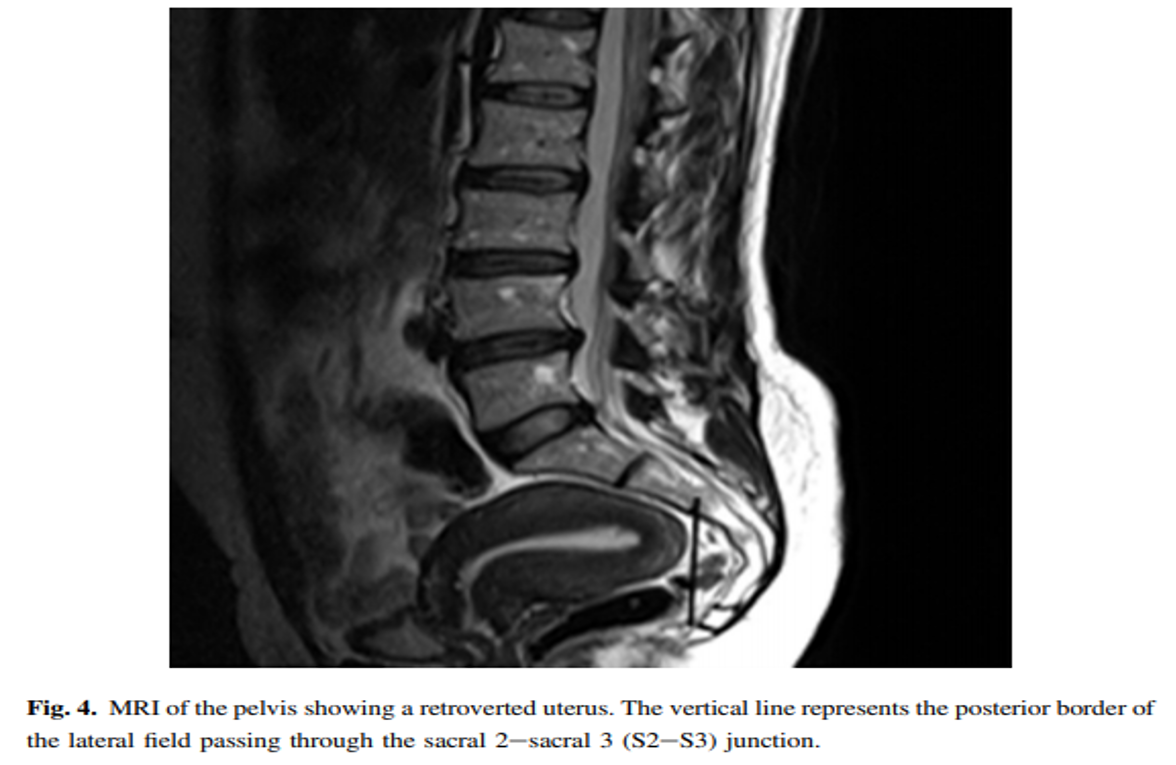
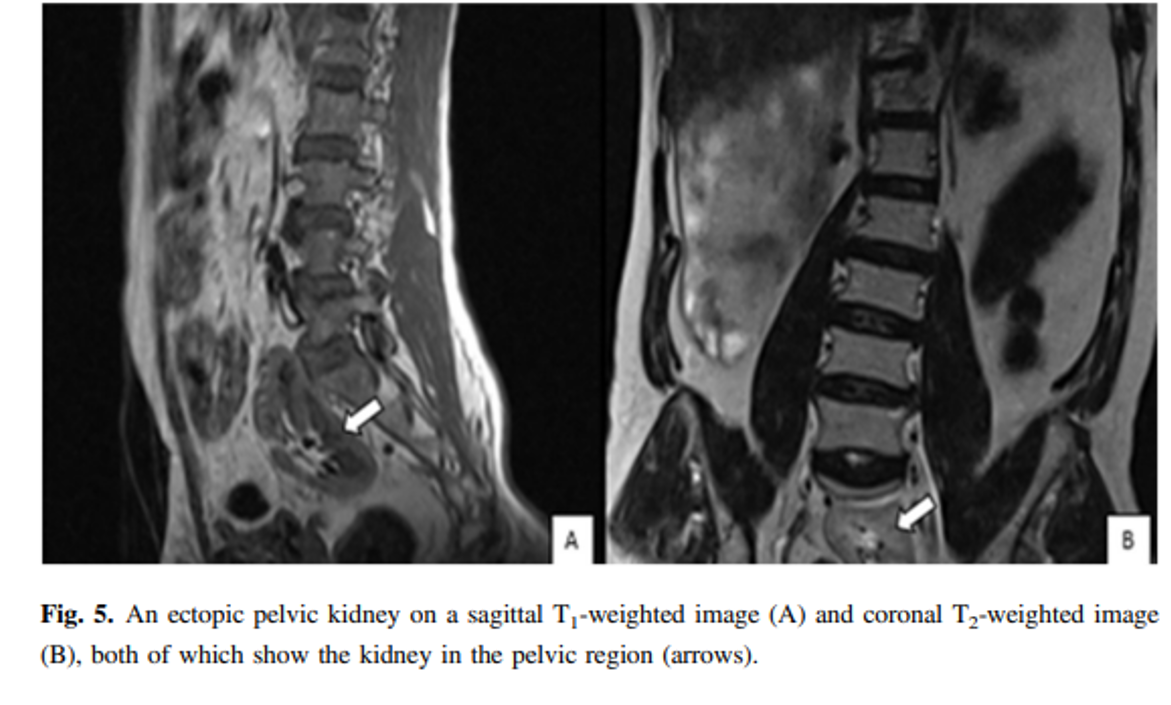
Results: Of the 379(100%) patients, 90(23.7%) patients were presented with incidental findings. Among the incidental findings, 39(10.3%) were renal cysts, 10(2.6%) were retroverted uteri, 5(1.3%) were Nabothian cysts, 4(1.1%) were ovarian cysts, 10(2.6%) were uterine fibroids, 3(0.8%) were endometrial thickening, 11(2.9%) were indicative of hydronephrosis, 4(1.1%) were uncovered prostatic enlargement, 2(0.5%) were atrophic kidney, and 1(0.3%) each was of an ectopic kidney and bladder wall thickening, respectively.
Conclusions: A high percentage of extraspinal pathological findings were detected during MRI lumbar spine scans of intervertebral discs. Thus, it is important to be aware of the high percentage of patients who undergo further evaluation given the presence of unexpected findings, but for whom clinical confirmation of these abnormalities is not obtained.
In this prospective study, a total of 379 patients-185(49%) males and 194(51%) females - were examined via lumbar spine MRI and presented with clinically suspected intervertebral disc diseases given the presence of low-back pain or sciatica symptoms. The common clinical features detected in these patients that were related to their complains were as follows: i) hip pain (n = 20; 5.3%); ii) burning or tingling sensation down the leg (n = 175; 46.2%); iii) weakness, numbness, or difficulty moving the leg or foot (n = 210; 55.4%); iv) pain in the rear or leg that is worse when sitting (n = 113; 29.8%); and v) a shooting pain that makes it difficult to stand (n = 320; 84.4%). The number and percentages of IESFs and congenital anomalies/anatomical differences presented during the lumbar spine MRI scans were 89(23.5%) and 1(0.3%), respectively (Table 1).
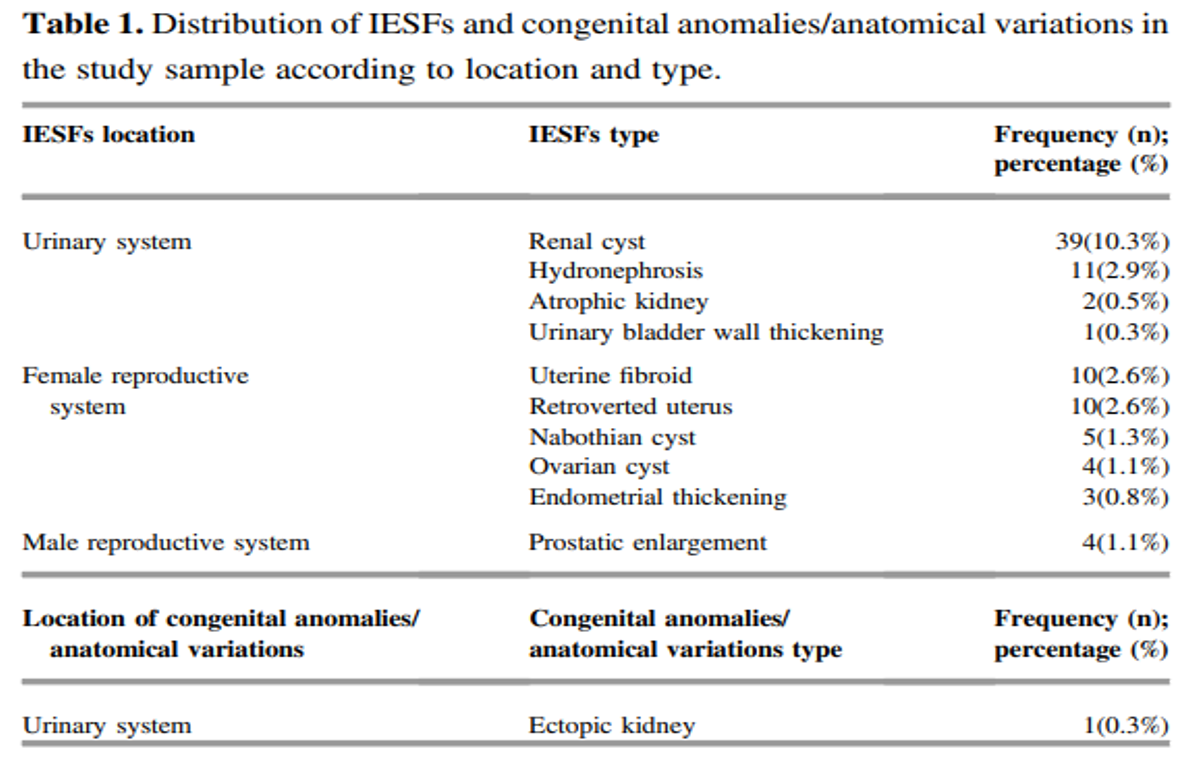
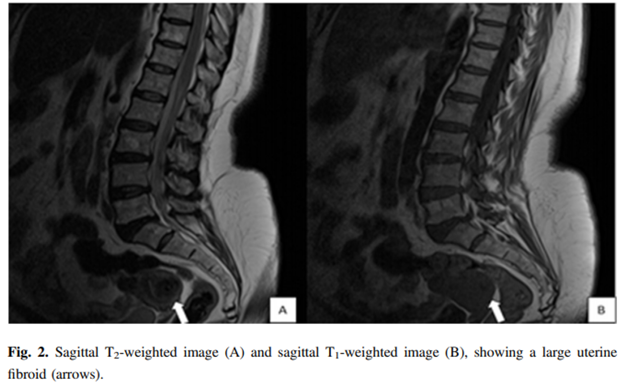
The incidence percentage of IESFs based on age groups were 2%, 7.5%, 17.9%, 35.8%, 28.3%, and 8.5% for age ranges 1-15 years, 16-30 years, 31-45 years, 46-60 years, 61-75 years, and 76 years, respectively. In addition, the IESFs were most likely related to the kidneys (n = 53; 13.7%) (Figs. 1 and 3) and were more likely to affect males (n= 4; 1.1%) and females’ (n = 32; 8.4%) internal genital organs (Figs. 2 and 4).
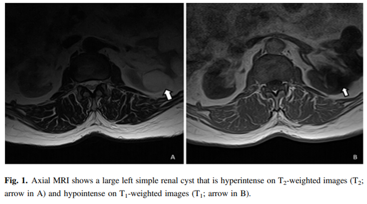
Renal cysts (n = 39; 10.3%) were the most common IESFs across MRI scans of the lumbar spine (Table 1 and Fig. 1). On MRI lumbar spine scans for female patients, uterine fibroids and a retroverted uterus were the most common IESFs, with incidence rates of 2.6% (n = 10) respectively (Table 1, Figs. 2 and 4).
Prostatic enlargement was detected as an IESFs in a total of 4 (1.1%) male patients on their MRI scans of the lumbar spine (Table 1). Only one case of ectopic kidney (0.3%) was registered as an incidental extraspinal congenital anomaly/anatomical variation as shown in Fig. 5. Although we identified these IESFs, congenital anomalies, and anatomical variations, further examinations and follow ups were not performed during this study; consequently, we did not obtain information on the outcome of these patients.
In conclusion, clinical judgement needs to be exercised when reporting IESFs, congenital anomalies or anatomical variations of the lumbar spine following MRI scans of this region, and guidelines are required to determine when further investigations are necessitated. In addition, IESFs, congenital anomalies, or anatomical variations are common in routine lumbar MRI scans, although their clinical significance is uncommon. Clinically significant IESFs are occasionally omitted from formal clinical reports. Along these lines, a methodical assessment of spinal and non-spinal structures in lumbar MR images may be of significance in clinical practice, as these images can have significant impacts on patient management and on the medicolegal ramifications to the radiologist.
The INCIDENTAL FINDINGS may be more significant than the problems being evaluated and can have significant impact on patient management and medicolegal implications to the radiologist.
The authors would like to thank Mr. Ahmed Fadlula Musa, Mr. Mohammed Hashim, Mr. Tarig Haga, Mr. Rafat Abdalla and Mr. Almoutasm Bellah, the staff of the MRI diagnostic centers of Al Zaytouna Specialist Hospital, Dar Al Elaj Specialized Hospital and El Nilein Medical Diagnostic Centre, Khartoum, Sudan, without whom this study would not have been possible. English-language editing of this manuscript was provided by Journal Prep Services.
[1] S.C. Wanger, W.B. Morrison, J.A. Carrino, M.E. Schweitzer, H. Nothnagel, Picture archiving and communication system: effect on reporting of incidental findings, Radiology 225 (2002) 500-505.
[2] C.C. Quattrocchi, A. Giona, A.C. Di Martino, Y. Errante, L. Scarciolla, C.A. Mallio, V. Denaro, B.B. Zobel, Extra-spinal incidental findings at lumbar spine MRI in the general population: a large cohort study, Insights Imag. 4 (2013) 301-308.
[3] C.Z. Liang, H. Li, Y.Q. Tao, X.P. Zhou, Z.R. Yang, F.C. Li, Q.X. Chen, The relationship between low pH in intervertebral discs and low back pain: a systematic review, Arch. Med. Sci. 8 (2012) 952-956.
[4] R. Chou, A. Qaseem, D.K. Owens, P. Shekelle, Clinical guidelines committee of the American College of Physicians, clinical guidelines committee of the American College of Physicians. Diagnostic imaging for low back pain: advice for high-value health care from the American College of Physicians, Ann. Intern. Med. 154 (2011) 181-189.
[5] F.A. Mettler Jr., B.R. Thomadsen, M. Bhargavan, D.B. Gilley, J.E. Gray, J.A. Lipoti, J. McCrohan, T.T. Yoshizumi, M. Mahesh, Medical radiation exposure in the U.S. in 2006: preliminary results, Health Phys. 95 (2008) 502-507.
[6] S.J. Golding, Radiation exposure in CT: what is the professionally responsible approach? Radiology 255 (2010) 683-686.
[7] S.Y. Lee, M.S. Landis, I.G. Ross, A. Goela, A.E. Leung, Extraspinal findings at lumbar spine CT examinations: prevalence and clinical importance, Radiology 263 (2012) 502-509.
[8] H.J. Park, Y.H. Jeon, M.H. Rho, E.J. Lee, N.H. Park, S.I. Park, J.H. Jo, Incidental findings of the lumbar spine at MRI during herniated intervertebral disk disease evaluation, AJR Am. J. Roentgenol. 196 (2011) 1151-1155.
[9] N. Magnavita, G. Magnavita, A. Fileni, A. Bergamaschi, Ethical problems in radiology: medical error and disclosure, Radiol. Med. 114 (2009) 1345-1355.
[10] M.A. Hlatky, C. Iribarren, The dilemma of incidental findings on cardiac computed tomography, J. Am. Coll. Cardiol. 54 (2009) 1542-1543.
[11] T. Xiong, M. Richardson, R. Woodroffe, S. Halligan, D. Morton, R.J. Lilford, Incidental lesions found on CT colonography: their nature and frequency, Br. J. Radiol. 78 (2005) 22-29.
[12] M.E. Zalis, M.A. Barish, J.R. Choi, A.H. Dachman, H.M. Fenlon, J.T. Ferrucci, S.N. Glick, A. Laghi, M. Macari, E.G. McFarland, M.M. Morrin, P.J. Pickhardt, J. Soto, J. Yee, CT colonography reporting and data system: a consensus proposal, Radiology 236 (2005) 3-9.
[13] D.K. Weiner, Y.S. Kim, P. Bonino, T. Wang, Low back pain in older adults: are we utilizing healthcare resources wisely? Pain Med. 7 (2006) 143e150.
[14] L. Berlin, The incidentaloma: a medicolegal dilemma, Radiol. Clin. North. Am. 49 (2011) 245-255.
[15] T. Sutherland, J.M.E. Chock, N. Stephens, Incidental extra spinal findings at CT lumbar spine on wide field of view reconstructions, J. Spine Neurosurg. 4 (2013) 4.
 Send Email