

Theme
Nuclear Medicine
INSTITUTION
King Fahad military medical complex / University of Western Ontario

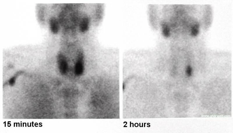
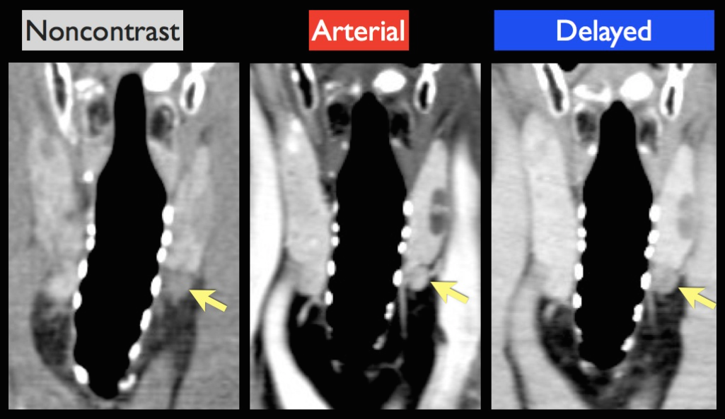
Accurate preoperative anatomic localization of hyperfunctioning parathyroid adenoma (HTP) critical importance in surgical planning for minimally invasive parathyroidectomy. SPECT/CT with 99mTc-sestamibi (99mTc-MIBI) important for detection of any metabolic active hyperfunctioning gland(s) localized in the neck or mediastinum. Ultrasonography (US) and Four-dimensional parathyroid CT (4D CT) increasingly offers potential advantages as an alternative primary investigation and is a common second-line investigation.
Retrospective study included 28 patients aged 26 - 87 years (mean age 49±7). Female to male ratio was 4:1. The follow-up period ranged from 3 to12 months, the mean being 6 months. In all cases of biochemically confirmed hyperparathyroidism, the average levels were: Ca total - 2.65±0.05mmol/l (NV 2.12-2,52), PTH=72±32pg/ml (NV 1.5-9.2). All underwent parathyroidectomy, and an adenoma was confirmed. Scintigraphy was performed with SPECT/CT (GE model 670pro NM/CT). 4D CT done with Toshiba aquilion 64. Ultrasonography (US) was done with ACUSON SC2000 by means of 10MHz linear probe.
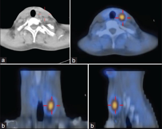
The exact localization hyperfunctioning parathyroid gland was identified 23 (82.2%) out of 28 patients by means of SPECT/CT. The remaining 5 patients need an alternative investigation and second-line investigation which we started with US parathyroid adenomia was identified 2 (40%) out of 5 patients. All remaining 5 patients further invastigated with 4D-parathyroid CT scan hyperfunctioning parathyroid gland was identified 4 (80%) out of 5 patients. One patients (3.5%) out of 28 patients was dignosed intraoperatively.
SPECT/CT with 99mTc-sestamibi appears promising with an important key role in diagnosis hyperfunctioning parathyroid gland but further imaging with 4D-parathyroid CT scan may needed particularly when we attempting to diagnose the precise site of disease localization in presurgical planning.
An integrated diagnostic approach based on scintigraphy and high-resolution neck ultrasound was extremely accurate of identifying patients with parathyroid lesions before surgery, enabling them to undergo limited surgery, 4DCT scan analysis may be useful in some patients with suspected parathyroid adenoma localised in a deep seat, allowing a more precise identification of the latter and better surgical planning.
1. Geatti, O . Parathyroid scintigraphy. Q J Nucl Med. 1999;43(3):207–216.
2. McBiles, M, Lambert, AT, Cote, MG, Kim, SY. Sestamibi parathyroid imaging. Semin Nucl Med. 1995;25(3):221–234.
3. Treglia, G, Sadeghi, R, Schalin-Jäntti, C. Detection rate of (99m) Tc-MIBI single photon emission computed tomography (SPECT)/CT in preoperative planning for patients with primary hyperparathyroidism: a meta-analysis. Head Neck. 2016;38(S1):E2159–E2172. doi:10.1002/hed.24027.
4. Wei, WJ, Shen, CT, Song, HJ, Qiu, ZL, Luo, QY. Comparison of SPET/CT, SPET and planar imaging using 99mTc-MIBI as independent techniques to support minimally invasive parathyroidectomy in primary hyperparathyroidism: a meta-analysis. Hell J Nucl Med. 2015;18(2):127–135. doi:10.1967/s002449910207.
5. Kluijfhout, WP, Vorselaars, WMCM, Vriens, MR, Borel Rinkes, IHM, Valk, GD, de Keizer, B. Enabling minimal invasive parathyroidectomy for patients with primary hyperparathyroidism using Tc-99m-sestamibi SPECT–CT, ultrasound and first results of 18F-fluorocholine PET–CT. Eur J Radiol. 2015;84(9):1745–1751. doi:10.1016/j.ejrad.2015.05.024.
6. Wong, KK, Fig, LM, Gross, MD, Dwamena, BA. Parathyroid adenoma localization with 99mTc-sestamibi SPECT/CT. Nucl Med Commun. 2015;36(4):363–375. doi:10.1097/MNM.0000000000000262.
7. Taywade, SK, Damle, NA, Behera, A. Comparison of 18F-Fluorocholine positron emission tomography/computed tomography and four-dimensional computed tomography in the preoperative localization of parathyroid adenomas-initial results. Indian J Endocrinol Metab. 2017;21(3):399–403. doi:10.4103/ijem.IJEM_536_16.
8. Hoang, JK, Williams, K, Gaillard, F, Dixon, A, Sosa, JA. Parathyroid 4D-CT. Otolaryngol Neck Surg. 2016;155(6):956–960. doi:10.1177/0194599816655311.
9. Hamidi, M, Sullivan, M, Hunter, G. 4D-CT is superior to ultrasound and sestamibi for localizing recurrent parathyroid disease. Ann Surg Oncol. 2018;25(5):1403–1409. doi:10.1245/s10434-018-6367-z.
10. Cheung, K, Wang, TS, Farrokhyar, F, Roman, SA, Sosa, JA. A meta-analysis of preoperative localization techniques for patients with primary hyperparathyroidism. Ann Surg Oncol. 2012;19(2):577–583. doi:10.1245/s10434-011-1870-5.
11. Kuzminski, SJ, Sosa, JA, Hoang, JK. Update in parathyroid imaging. Magn Reson Imaging Clin N Am. 2018. doi:10.1016/j.mric.2017.08.009.
12. Mahajan, A, Starker, LF, Ghita, M, Udelsman, R, Brink, JA, Carling, T. Parathyroid four-dimensional computed tomography: evaluation of radiation dose exposure during preoperative localization of parathyroid tumors in primary hyperparathyroidism. World J Surg. 2012;36(6):1335–1339. doi:10.1007/s00268-011-1365-3.
13. Rausch, I, Füchsel, FG, Kuderer, C, Hentschel, M, Beyer, T. Radiation exposure levels of routine SPECT/CT imaging protocols. Eur J Radiol. 2016;85(9):1627–1636. doi:10.1016/j.ejrad.2016.06.022.
14. Nael, K, Hur, J, Bauer, A. Dynamic 4D MRI for characterization of parathyroid adenomas: multiparametric analysis. AJNR Am J Neuroradiol. 2015;36(11):2147–2152. doi:10.3174/ajnr.A4425.
15. Merchavy, S, Luckman, J, Guindy, M, Segev, Y, Khafif, A. 4D MRI for the localization of parathyroid adenoma: a novel method in evolution. Otolaryngol Head Neck Surg. 2016;154(3):446–448. doi:10.1177/0194599815618199.
16. Caldarella, C, Treglia, G, Isgrò, MA, Giordano, A. Diagnostic performance of positron emission tomography using 11C-methionine in patients with suspected parathyroid adenoma: a meta-analysis. Endocrine. 2013;43(1):78–83. doi:10.1007/s12020-012-9746-4.
17. Krakauer, M, Kjaer, A, Bennedbæk, F. 18F-FET-PET in primary hyperparathyroidism: a pilot study. Diagnostics. 2016;6(3):30. doi:10.3390/diagnostics6030030.
18. Lezaic, L, Rep, S, Sever, MJ, Kocjan, T, Hocevar, M, Fettich, J. 18F-Fluorocholine PET/CT for localization of hyperfunctioning parathyroid tissue in primary hyperparathyroidism: a pilot study. Eur J Nucl Med Mol Imaging. 2014;41(11):2083–2089. doi:10.1007/s00259-014-2837-0.
19. Kluijfhout, WP, Pasternak, JD, Drake, FT. Use of PET tracers for parathyroid localization: a systematic review and meta-analysis. Langenbecks Arch Surg. 2016;401(7):925–935. doi:10.1007/s00423-016-1425-0.
20. Cassinello, N, Ortega, J, Lledo, S. Intraoperative real-time 99mTc-sestamibi scintigraphy with miniature gamma camera allows minimally invasive parathyroidectomy without ioPTH determination in primary hyperparathyroidism. Langenbecks Arch Surg. 2009;394(5):869–874. doi:10.1007/s00423-009-0523-7.
 Send Email