


Theme
Radiologic Technology
INSTITUTION
The National Ribat University,Faculty of Radiological and Nuclear Medicine Science

Lower back pain can best described in terms of specific accompanying features, it can be caused by a variety of problems with any parts of the complex, interconnected network of spinal muscles, nerves, bones, discs or tendons in the lumbar spine [1].
The lumbar (or lower back) region is made up of five vertebrae (L1-L5), sometimes including the sacrum. In between these vertebrae are fibro cartilaginous discs, which act as cushions, preventing the vertebrae from rubbing together while at the same time protecting the spinal cord.
Nerves come from and go to the spinal cord through specific openings between the vertebrae, providing the skin with sensations and messages to muscles. Stability of the spine is provided by the ligaments and muscles of the back and abdomen. Small joints called facet joints limit and direct the motion of the spine [2].
The lumbar spine series is comprised of two standard projections along with a range of additional projections depending on clinical indications. The series is often utilized in the context of trauma, postoperative imaging and for chronic conditions such as ankylosing spondylosis [2].
Lumbar spine x rays are the most commonly ordered radiographic investigation of the spine, however, it is widely documented that plain radiography is far inferior in the investigation of suspected lumbar spine pathology compared to that of MRI and CT. Although lumbar spine x-rays are a part of general back pain workups there is no evidence that obtaining x rays before other modalities will result in higher patient outcomes [2].
The aim of this study is to evaluate lower back pain in soldier patients using conventional x- ray.
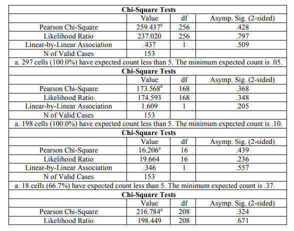
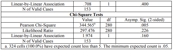
This descriptive cross sectional study, aimed to evaluate the lower back pain using conventional x-ray in soldiers patients. The problem of this study was no similar study done on this topic and no reference value for it. The study was done in 153 patients in range of age between 27-65 years came for back x-ray. The study was done in Ribat Hospital University in period from December 2017 to May 2018. The data was collected and analyzed by statistical package for social science and correlations person's coefficient. The study shows common causes of lower back pain were; 15.7%, Spondylitis 11.1%, multiple disc 11.1%, loss of lordosis 12.4%, hyper lordosis 7.8%, lumbar scoliosis 5.2%, compression fracture 8.5%, and spondylolysis 6.5%. There was significant relation between working years of soldier and x-ray findings and less significant relation between working place of soldier and x-ray findings (p=0.671, p=0.348) respectively.
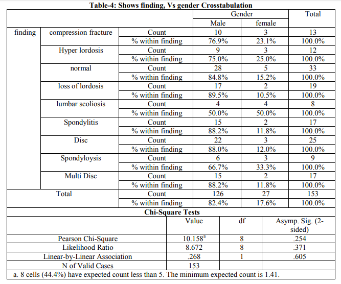
In this study, x-ray findings in patients with pain limited to their lower back pain were analyzed .Regarding frequency distribution of patients under study according to gender, there were 126 males (82,4%) and 27 females (17,6%). This matches with study done by Andrew P kant, Wayne J Daum S Michael Dean, they found that male (79%) are more affected than female (21%) [3].
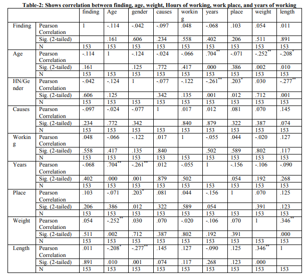
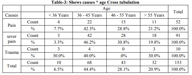
According to age frequency distribution of patients under study, it was categorized into four groups, group (A) less than 35 was 10 patients (6.5%), group (B) were from 36 to 45 was 68 patients (44.4%), group (C) from 46 to 55 was 43patients (28%), group (D) were more than 55years old, they was 32 patients (20.9%), from this we found that the most affected age was group(B) their age from 36 to 45 years , this was matches with Mohd Nazeer who found that the most affected age between 31 to 40 years old [3].There is less significant relation between age and finding ( p = 0 .0226) [4].
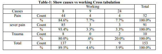
According to hours of working, the frequency distribution of patients under study was categorized into three groups (A) the working hours were 8 hours, was 137 patients (89.5%), group (B) the working hours was 9 hours, were 7 patients (4.6%) and group (C) the working hours was 24, this was 9 patients (5.9%), not found in previous study, there is less significant relation between working and finding (p = 0.0236).
According to causes of lower back pain, frequency distribution of patients under study was categorized into three groups; group (A) patients for pain at 52 patients (34%), group (B) patients for severe pain at 91 patients (59.5%), and group (C) patients for trauma at 10 patients (6.5%). According to x-ray finding frequency distribution of patients under study, it was categorized into nine groups, group (a) patients with compression fracture is 13 patients (8.5%), group (b) patients with hyper lordosis is 12 patients (7.8%), group (c) normal patients 33 patients (21.6%), group (d) patients with loss of lordosis 19 patients (12.4%), group (E) patients with lumber scoliosis 8 patients (5.2%), group (f) patients with spondylolysis 17 patients(11.1%), group (g) patients with disc 24 patients(15.7%), group (h) patients with spondylolysis 10 patients (6.5%), group (i) patients with multiple disc 17 patients (11.1%). According to x-ray findings and lumbar spine effected frequency distribution of patients under study, it was categorized into five groups, group(a) effected from L1to L2 was 28 patients (18.3%),and the same in group (b), group (c) were from L3 to L4 was 34 patients (22.2%), group (d) to L4-L5 was 22 patients (14.4%), group (e) were from L5-S1 was 41 patients (26.8%), from this we found that the most effected spine level (L5 to S1) this was match with by: Andrew p Kant [4].
According to weight frequency distribution of patient under study, it was categorized into fife groups, group (A) were from 51 to 60 was 4 patients (2.6%), group (B) were from 61 to 70 was 46 patients (30,1%), group (C) were from 71 to 8o was 73 patients (47.7%), from this we found that the most effected ( 71 to 80 ) weight, this was no matches, group (d) were from 81 to 90 was 25 patients (16.3%), group (e) more than 90 was 5 patients (3.3%), there is strong significant relation between weight and finding. In this study we found more significant relation between years of service and finding (P = 0 .0671). There is less significant relation between working place and finding (p = 0 .0348).
Lower back pain is common in soldier's patients; obviously, they have relation with age, weight and length. The analysis of x-ray findings in this study showed that Spondylitis, loss of lordosis, hyper lordosis, spondylolysis, lumbar scoliosis, disc, multiple disc are common in soldiers patients. Disc prolapse is common causes of lower back pain 15.7% of this study. The common level of lumbar spine affected by disease is L5-S1in the study.
Before doing x-ray, complete history is necessary to determine the position and factor for radiological examination. The important educational points for patients with lower back pain are necessary. More research should be done using a larger sample of patients for further assessment.
1. Kerry H. Levin MD. August Low back pain, The Cleveland clinic foundation, Center for Continuing Educating. 2010.
2. Neill A. Back pain 29. Challenging Concepts in Emergency Medicine: Cases with Expert Commentary. 2015 Mar 19:285.
3. Balagué F, Mannion AF, Pellisé F, Cedraschi C. Non-specific low back pain. The Lancet. 2012 Feb 4;379(9814):482-91.
4. Hoy D, March L, Brooks P, Blyth F, Woolf A, Bain C, Williams G, Smith E, Vos T, Barendregt J, Murray C. The global burden of low back pain: estimates from the Global Burden of Disease 2010 study. Annals of the rheumatic diseases. 2014 Jun 1;73(6):968-74.
 Send Email