
| Theme: Radiologic Technology | |||
 |
||||||
| The Effect of Manual Shimming on MR Spectrum Quality |
 |
|||||
|
||||||

Magnetic Resonance Spectroscopy (MRS): Is an analytical in vivo chemical method that enables the identification and quantification of metabolites in samples which is a noninvasive method compare to other producers such as biopsy (Bertholdo et al., 2013). New developments in Magnetic Resonance Imaging (MRI) scanners technology made MRS a wildly used technique. Despite these recent improvements in technology, performing MRS in clinical sitting still face some challenges of B0 field Inhomogeneity in certain areas in the brain (figure 1) determines the spectral resolution of the MRS result (Lin et al., 2012).
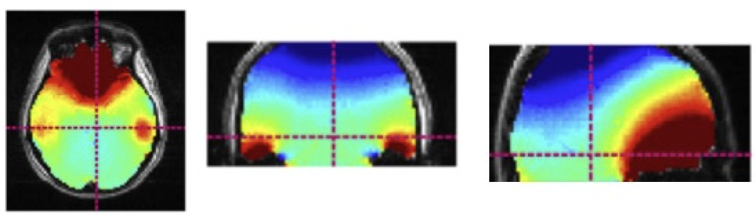
Figure 1: Whole-brain B0 homogenization in auto-shimming (Juchem , 2016, p.9)
These inhomogeneities in the human brain can be explained by the susceptibility differences between air and tissue. Spectral resolution is one of the most important factors to evaluate any giving MRS experiment. Acquisitions with a low spectral resolution may lead to inaccurate metabolite quantification (Image below). Improved magnetic field homogeneity increases SNR and narrows peak widths-thus improves both sensitivity and spectral resolution. Spectral resolution represents peaks width, which determines whether metabolites under investigation can be separated. Better filed homogeneity can be achieved by second or third-order shimming (also known as manual shimming). This project's aim is to evaluate the effect of manual shimming on spectral quality in clinical settings (Lin et al., 2012 and Blüml, 2013).
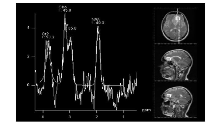
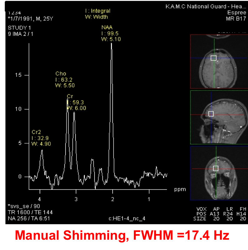
MRS of a patient with frontal lobe lesion by KAMC, 2015
All imaging and spectroscopy were performed at 1.5 Tesla using a standard clinical MR scanner, head coil, and software (MAGNETOM® Espree; Siemens Medical Systems) operated and maintained under the manufacturer's specifications.
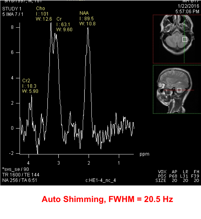
1- First scan (Automatic shimming):
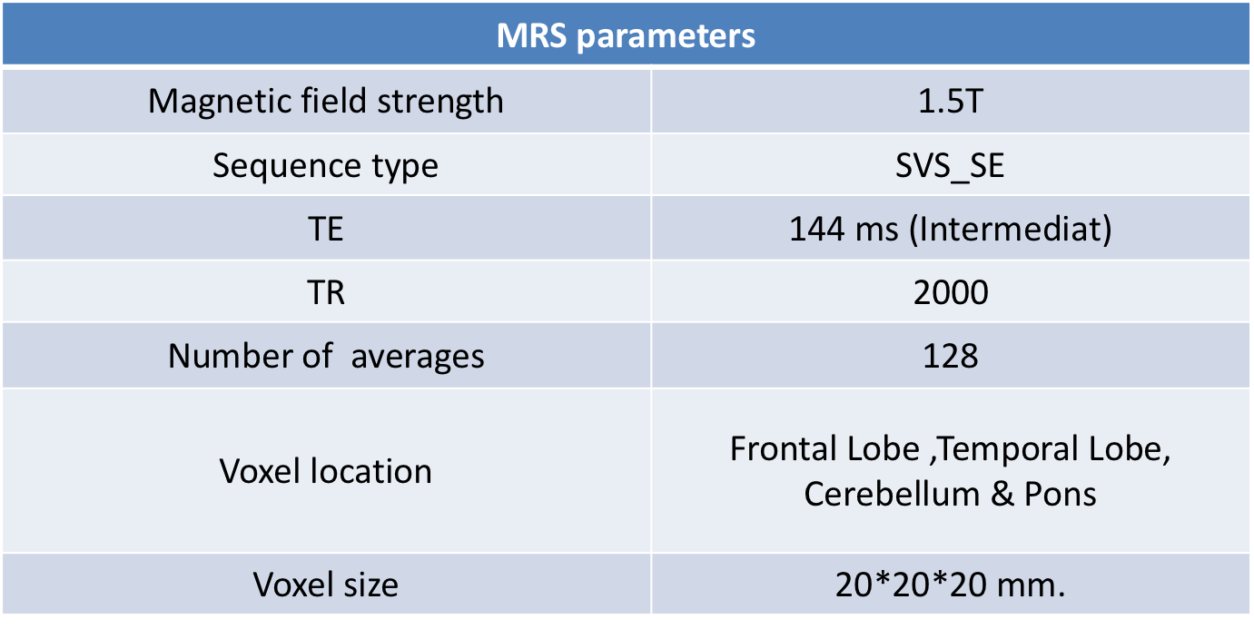
Voxels of single‐voxel spectroscopy (SVS) point‐resolved spectroscopy (PRESS), (TE = 140ms, TR = 2000 ms) were prescribed using the axial, sagittal and coronal T2W images as references. SVS PRESS spectra were acquired at the Frontal lobe, Temporal lobe, Pons and Cerebellum levels. Automated optimization of gradient shimming, transmitter pulse power, and water suppression was used. The total acquisition time was 5 min per spectrum. Detailed MRS pulse parameter in the table below.
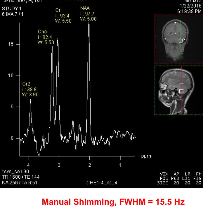
2- Second scan (Manual shimming):
The same single‐voxel spectroscopy was repeated applying manual shimming as it will be explained in (figure 2).
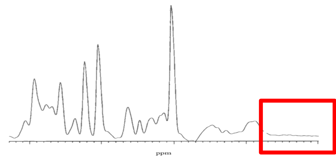
• The following criteria were used to evaluate spectrum quality:
1.Peak FWHM (figure 3).
2. Signal adequately higher than noise?
3.Are peaks sufficiently narrow?
- Can Creatine and Choline be adequately identified?
4.Did water suppression work?
5.Did all metabolites clearly appear?
6. Baseline is rarely flat from 0.8 – 1.5 ppm
- Manual Shimming Steps
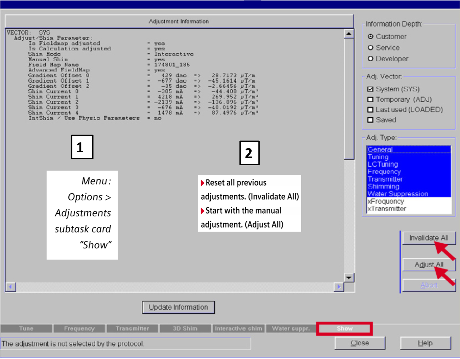
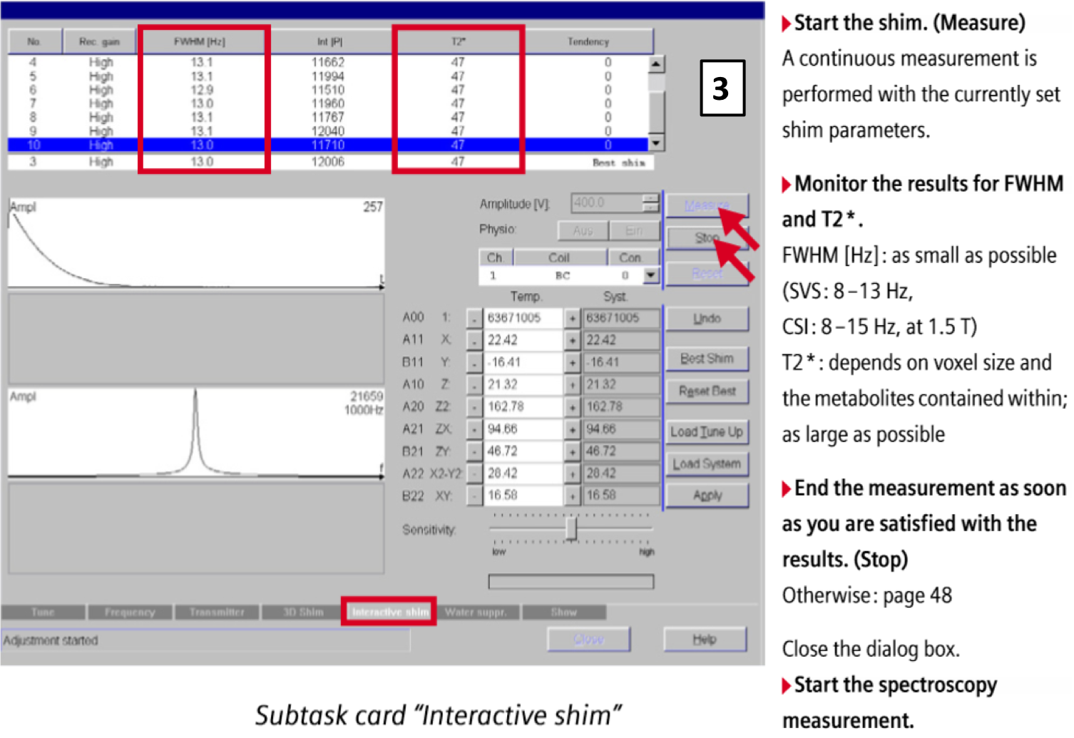

Figure 2: Adjust manual shimming (Siemens, 2009, p.25-28)
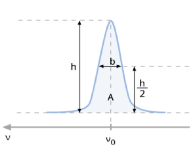
vo: Resonance frequency
h: Height of peak
b: Full Width at Half Maximum, FWHM
A: Integral
Figure 3: Characteristics of a peak (Siemens, 2010-2012, p.16)
- 4 participants included in the study. A total of 8 MRS experiments were done. The average time to perform manual shimming was 7 minutes (min=5, max =13).
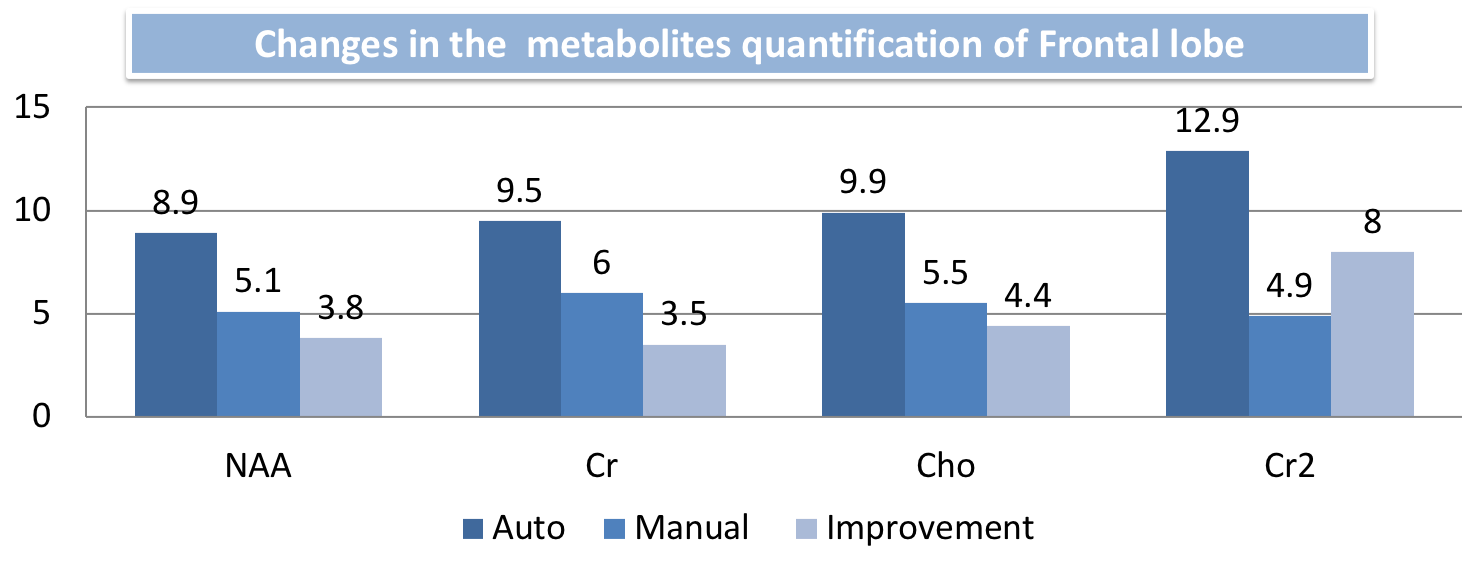
- The most significant improvement was in the frontal lobe and Cerebellum.
- Area near to the base of the skull was the most challenging area to perform manual shimming.
Frontal lobe

.
Cerebellum
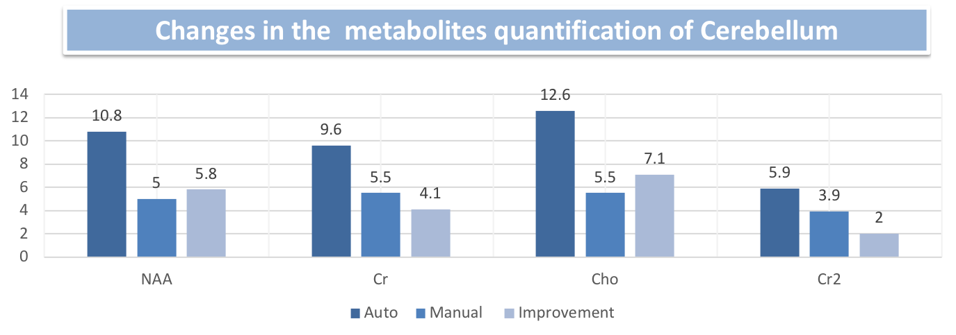
- The table below shows all results:
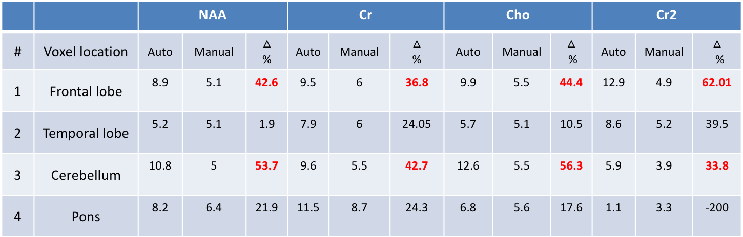
- Manual shimming can significantly improve MR spectrum quality, especially in difficult areas.
- Based on this experiment manual shimming should be conducted with multivoxels spectroscopy because of highly inhomogeneity.
• Limitations:
1- Small sample size.
2- Experiment done just in one machine “Siemens”.
•Figure 1: Juchem, C, Graaf, R.A. de. 2016. B0 magnetic field homogeneity and shimming for in vivo magnetic resonance spectroscopy. Analytical Biochemistry. 529, pp17-29.
•Figure 2: Syngo MR B15VOperator Manual – Spectroscopy. 2009. Starting protocol adjustments. [online]. [Accessed by Jan 2016]. Available from: https://www.siemens-healthineers.com/magnetic-resonance-imaging/magnetom-world/clinical-corner/application-tips/syngo-mr-b15v-operator-manual-spectroscopy.html
•Figure 3: Syngo MR D13 Basic Manual – Spectroscopy. 2010-2012. Characteristics of a peak. [online]. [Accessed by Jan 2016]. Available from: https://cbbi.udel.edu/wp-content/uploads/2017/01/BMSpectroscopy_Eng.pdf
•Bertholdo, D, Watcharakorn, A, Castillo, M. 2013. Brain Proton Magnetic Resonance Spectroscopy: Introduction and Overview. Neuroimaging Clinics of North America. 23(3), pp 359-380
•Lin, A, Tran, T, Bluml, S, Merugumala, S, Liao, HJ, Ross, BD. 2012. Guidelines for acquiring and reporting clinical neurospectroscopy. Semin Neurol, 32(4), p432-453.
•Blüml S. 2013. Magnetic Resonance Spectroscopy: Basics. In: Blüml S., Panigrahy A. (eds) MR Spectroscopy of Pediatric Brain Disorders. Springer, New York, NY, pp11-23.
 Send Email