


Theme
Medical Physics
INSTITUTION
1 Industrial Research and Consultancy Center (IRCC)
2 Radiation and Isotopes Center Khartoum (RICK)
Breast cancer is the most frequent women malignancy over the world, cause in enormous mortality rate (1). Computational radiation dosimetry has been used prominently in organs and tissues dose computations follow on breast imaging and radiotherapy techniques (2, 3), these studies have need of a computational models as a data for Monte carlo simulation.
The first generation of a computational models was the Mathematical which has limitations respect to its competence to describe complexity and reality of the human anatomy, it's problem was solved with the more reliable second generation Voxel, recently hybrid phantom was introduced, based on a poly mesh surfaces and Non Uniform Rational Spleen (NURBS) combining the flexibility of the mathematical and realism of voxel, but it's deferomation still difficult process and needs some efforts and cutting edge specialists. Depending on these types using diverse methods, dedicated Computational phantoms has been modeled for the Breast showed the positive reflection to adress problems (4, 5).
In this work we developed a method to obtain an artificial tomographic data as solution to simplify accessing data for Breast computaional dosimetry studdies, verification was done by Gate/Geant4 Monte Carlo code.
Preparing an easy adapted tommographic breast data was the main issue, which achieved through the following main processes, starting point was preparation of the surface models, Make HumanTM 3D software it’s (GUI) appears in figure 1a , was used to get the outer surface poly mesh models with desired breast features, three breast sizes were chosen; A, C and E according to the European standarization system of the dress. The surface models was exported to BlenderTM software it’s GUI can be seen in figure 1a, where the modification of the outer surfaces and placement of the internal organs skeleton, heart and lungs in their correct anatomical positions was done, the results were poly mesh chest models easy to manipulate as shown in figure 1c. The prepared poly mesh surface models were converted to voxel ones as illustrated in figure 1d using Binvox software. Every voxel model was created using Binvox in matrix size of 256*256*256 and voxel dimension of 1.4*1.4*1.4 mm3. MATLAB® was used to reslice the models axially into 5 mm and the segmentation was done into four body regions heart, lungs, bone and soft tissue as shown in figure 1e.
The following process was using th Gate Monte Carlo code to study the effect of breast size on dose distribution. Irradiation of left breast virtual tumor bed was done for the breast models in supine position using a plane of 5*5 cm2 with normal incidence electron therapeutic beams havings energies of 6, 12 and 18 MeV, the geometry of the phantom appears in figure 1f.
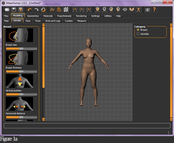
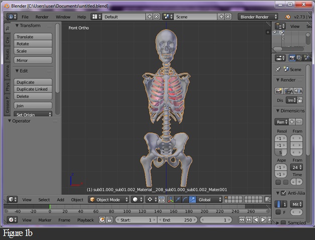
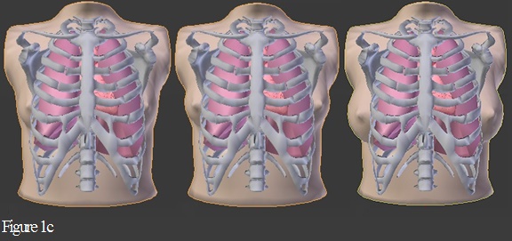
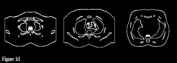
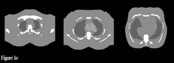
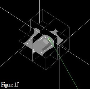
Figure1. Represent the main process to prepare the tommographic data
A colored map absorbed dose distribution in the central axial tommographic slices of the beams normalized to maximum in each of the small, medium and large breasts for 6, 12 and 18 MeV electron beams which represented in figures 2a, 2b and 2c respectively. Targeting (virtual tumor bed) 2.5 cm in distant from chest wall. Distribution within the different breast sizes explain the effect of the anatomical shape and size of the breast. 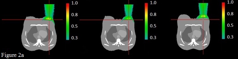
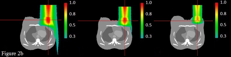
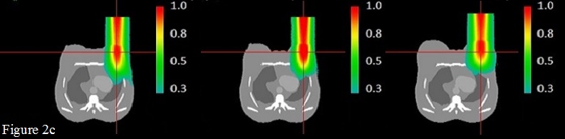
Figure 2. Verification of the usability the tommographic data for radiation computational dosimetry
The results of Gate Monte Carlo simulation seems to be in good similarity with known practice in radiotherapy, they proved efficiency of this method and tommographic data produced for computational dosimetry studies. The presented challenge is to apply this system in the area of research to solve different problems.
1. Jemal A, Bray F, Center MM, Ferlay J, Ward E, Forman D. Global cancer statistics. CA Cancer J Clin. 2011;61(2):69-90.
2. Yi Y, Lai CJ, Han T, Zhong Y, Shen Y, Liu X, et al. Radiation doses in cone‐beam breast computed tomography: A Monte Carlo simulation study. Medical physics. 2011;38(2):589-97.
3. Berris T, Mazonakis M, Stratakis J, Tzedakis A, Fasoulaki A, Damilakis J. Calculation of organ doses from breast cancer radiotherapy: a Monte Carlo study. Journal of applied clinical medical physics. 2013;14(1):133-46.
4. Dang J, Lasaygues P, Zhang D, Tavernier S, Felix N, et al. Development of breast anthropomorphic phantoms for combined PET-Ultrasound elasto- graphy imaging. IEEE International MedicalImaging Conference (2009) 3088–3092.
5. D. R. Dance, R. A. Hunt, P. R. Bakic, A. D. A. Maidment, M. Sandborg, G. Ullman, G. A. Carlsson, “Breast dosimetry using high-resolution voxel phantoms,” Radiat. Prot. Dosim., 114, 359–63 (2005).
I would like to express my very great appreciation to Dr. Elhussein Sirelkhatiem for his valuable supervision, my grateful thanks also extended to my family and friends for them support.
Special thanks should be given to the International Conference Radiation Medicine (ICRM) scientific and Organising committe for availing this opportunity.
 Send Email