
 |
Title
Integration of 3D Virtual Reality (VR) in Diagnostic and Therapeutic Imaging

Theme
Diagnostic & Interventional Radiology

Background
|
|
 |
Aim
To design an integrated platform that reads DICOM images from the scanner as an input and generates an interactive 3D virtual model of the scanned organ.
Summary of Work
|
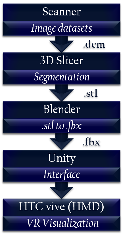
Scanner Medical image data sets from imaging modalities such as CT and MRI are loaded on to 3D Slicer as DICOM (Digital Imaging and Communications in Medicine) files.
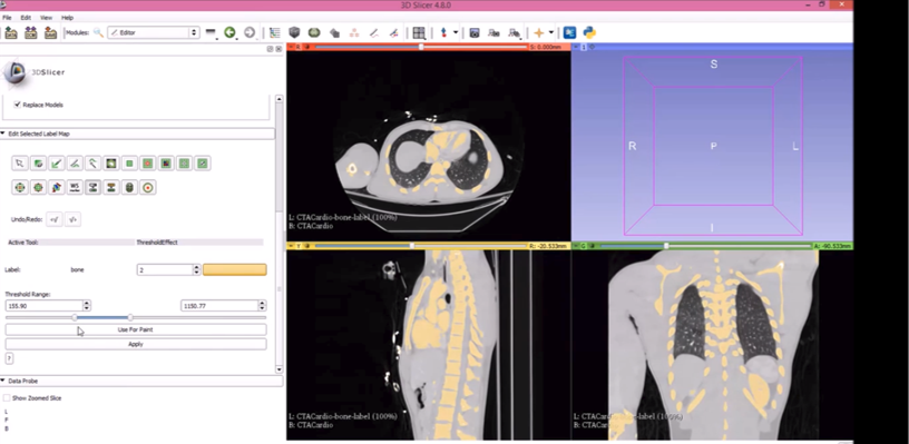
3D Slicer Using the image registration toolkit (ITK) in 3D Slicer, threshold based segmentation is performed. The threshold is chosen based on the range which best covers the bone tissue. This was 155.90 - 1150.77 for the CTA scan chosen in the trial. After each slice in the dataset has been segmented, the 3-dimensional volume model is rendered. This model is then exported in an .stl format as a surface rendered model.
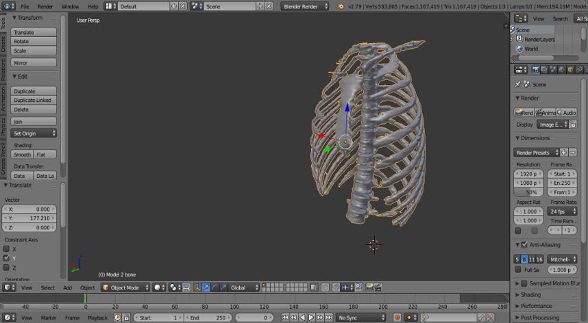
Blender As Unity does not provide direct support for stl data format, the surface render is converted from .stl to .fbx using Blender, which has support for both formats. Next, the .fbx file is imported to Unity where it is interfaced with the Vive.
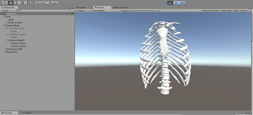
Unity A steamVR plugin is downloaded from the Unity asset store. This allows for placement of camera, and controller rigs that allows the scene to be viewed and interacted using the Vive. Each object is placed such that there is an appropriate view.
Head-mounted Display The scene is played and the view transfers to the VR head-mounted display (HMD). The user can view the rendered model in virtual reality and interact with it using the controllers. |
|
|
Summary of Results
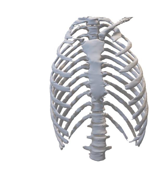
We were able to obtain a surface render model of the CTA 3D image dataset. The surface render can be virtually viewed by the HTC Vive HMD. At present, the user is only able to view the surface rendered model.
 |
 |
|
3D Slicer (Segmentation)
|
Surface Render product from 3D Slicer
|
 |
 |
|
Blender to convert image from .stl to .fbx |
Unity to interface with the Head-mounted VR headset |
Conclusion
Technology is advancing at a rapid rate. With VR integrated into medical imaging, efficient and accurate diagnosis could be made leading to better healthcare. Finer details may be viewed and an intuitive feeling of the scanned organ will be gained by the specialist which will make it easier to plan patient prognosis.
FUTURE WORK
The project is still ongoing and we are working to achieve the following:
- Ability to interact with the model
- Obtaining a volume render instead of surface render to obtain information from each voxel of the model
- Focusing on the coronary artery for quantitative and qualitative assessment of plaque formation
Take-home Messages
Improving the efficacy of medical imaging through Virtual Reality
- Detailed 3D realistic and interactive visualization will allow accurate diagnosis
- It will assist in the planning of complicated surgical procedures
- It has the potential to enhance medical education
References
[1] F. King, "An immersive virtual reality environment for diagnostic imaging," Dissertation/Thesis, ProQuest Dissertations Publishing, 2015.
[2] J. Egger, M. Gall, J. Wallner, P. Boechat, A. Hann, X. Li, X. J. Chen, and D. Schmalstieg, "HTC Vive MeVisLab integration via OpenVR for medical applications," PLOS ONE, vol. 12, p. e0173972, 2017.
[3] Virtual Reality in Medicine: New Opportunities for Diagnostics and Surgical Planning. Available: https://www.unibas.ch/en/News-Events/News/Uni-Research/Virtual-Reality-in-Medicine.html