
| Theme: Clinical Sciences 3 | |||
 |
||||||
| The Role of Sarcolemmal Membrane Associated Protein (SLMAP) in the Regulation of Cardiac Remodeling. |
 |
|||||
|
||||||

Heart diseases are still the most common causes of morbidity and mortality worldwide. Cardiac remodeling (increase and change in heart mass and shape) occurs naturally during development however, pathological remodeling starting with myocardial hypertrophy and progressing through dilated cardiomyopathy DCM adversely affects cardiac function often leading to sudden death. It is well established that calcium overload results in pathological remodeling of the myocardium however the therapeutic strategies to salvage the diseased myocardium is not completely successful. The sarcolemmal membrane associated protein (SLMAP-1, -2, -3) were first characterized in cardiac membrane and contribute to cardiac cell function. They were reported to regulate intracellular [Ca2+]i in the hearts well as cell proliferation, yet their role in cardiac remodeling is not yet elucidated. In this study we aimed to investigate the role of SLMAP in pathological remodeling by using Spontaneously Hypertensive Rats (SHR) and human DCM hearts (post-mortem). Our data provides new insight on the role of SLMAP in remodeled myocardium and cell proliferation.
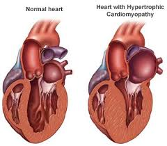

Fig 1: A normal heart is shown on the left compared to a heart with a hypertrophic cardiomyopathy (middle): there is obvious increase in the thickness of the walls of the left ventricle. The heart on the right is dilated: there is increased dimensions of the left ventricle and thinning of the LV wall. this figure was adopted from the mayo foundation for medical education and research
The models used were:
1- Proteins from LV of human hearts (*) from patients with dilated cardiomyopathy (DCM)- Post mortem.
2- Hearts from Spontaneously Hypertensive Rats (SHR) and Wistar-Kyoto rats (WKY) (*) with different ages 4-, 14-, and 18-week-old (to monitor the expression of SLMAP during the onset and progression of pathological remodeling.
(*) Handling of human and animal tissues was approved by the Ethics Committee and the Animal Care Committee at King Faisal Specialist Hospital and Research Center.
Hearts were harvested and dissected into left and right ventricles (LV and RV) and left and right atria (LA and RA).
Proteins were extracted and separated by western blotting (immunoblotted with specific SLMAP antibody).
Knockdown of SLMAP-3 was employed in Hela cells to monitor cell proliferation/migration using the proliferation assay kit (MTT)
The expression of SLMAPs is altered in the atria of SHR and WKY hearts
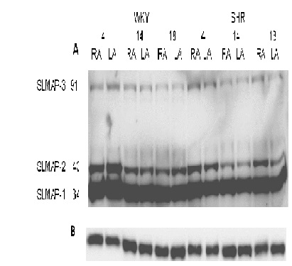
Figure 2:
A- Expression pattern of SLMAPs in right and left atria (RA and LA) of SHR and WKY hearts at 4, 14, and 18 weeks of age. The highest expression is noticed at 4 week
B- GAPDH of WKY and SHR
SLMAP-2 was higher in both RA and LA of WKY and SHR hearts
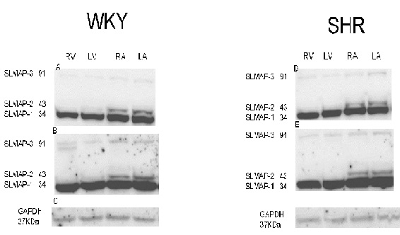
Figure 3: Expression level of SLMAP isoforms in WKY & SHR.
A- WKY 14week B- WKY 18week C- GAPDH for WKY 18week
D- SHR 14week E- SHR 18week F- GAPDH for SHR 18week
A novel SLMAP isoform occurs below the 91 KDa SLMAP3 in LV of SHR
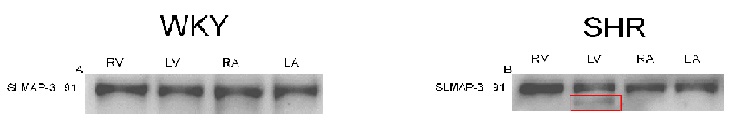
Figure 4: expression of SLMAP-3 isoform in WKY & SHR
A- SLMAP-3 in WKY at the age of 4 weeks.
B- SLMAP-3 in SHR at the age of 4 weeks (note: the appearance of a novel isoform of slmap-3 in the LV of SHR hearts)
SLMAP3 is lower in DCM LV compared to Normal LV
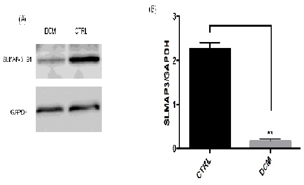
Figure 5: SLMAP3 expression is reduced in human DCM hearts
(A)Western blot of human LV hearts from DCM patients (post-mortem) showing a significant loss of SLMAP3 expression in DCM hearts compared to normal subjects.
B)Densitometry analysis of SLMAP3 bands using image J software **p<0.05 using T-test
Silencing SLMAP3 gene reduced cell growth/proliferation
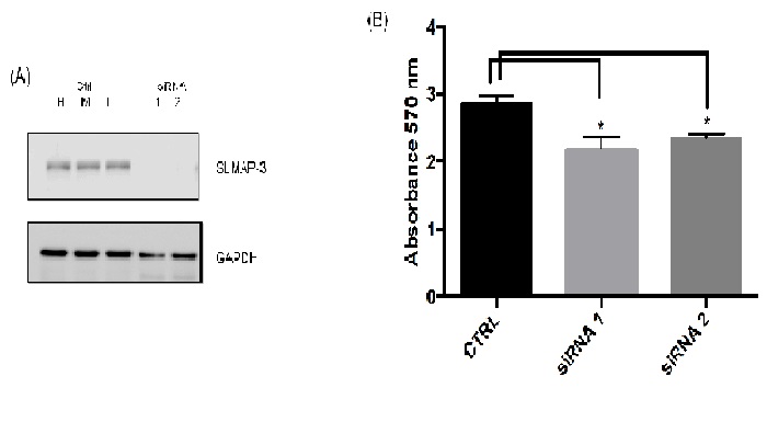
Figure 6: Data showing growth and proliferation compared between the cells transfected with SLMAP siRNA and control siRNA (CTRL) for 48 hour. *P<0.05 using T-test.
Our preliminary results indicate that SLMAP isoforms are regulated during cardiac remodeling in both SHR and DCM hearts. SLMAP-3 was highly expressed at 4 weeks and gradually decreased at 14- and 18- weeks of age. In human DCM, SLMAP3 was lower relatively to WKY. We also showed that the a novel band below the 91 KDa SLMAP3 (~72 KDa) appeared in the LV of SHR. Our data also indicate that SLMAP3 is implicated in the regulation of cell growth/proliferation.
This work is supported by a KACST operating grant (#13-BIO1452-20) to MN and PK, and by Alfaisal University IRG
 Send Email